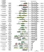

“4Peaks” was used to view and edit the sequence trace files ( ). The left half of Fig 3 shows the amplification curves of Eprobe-PCR, and the right half shows the electrogram of Sanger sequencing. The data were then transferred to Microsoft Excel (Microsoft, Redmond, WA, USA) and Cp values were evaluated.Įvaluation of the nine mutant tumors detected by Eprobe-PCR.Ĭomparison of three genotyping methods in all clinical samples.Ĭomparison of Eprobe-PCR and Sanger methods.

(c) Cp (crossing point) values of two experiments (a) and (b) were calculated by the second derivative maximum method in the LightCycler480. Traces appear sharper when viewed through 4Peaks compared to other chromatogram viewing programs, which allows for a easy viewing. The light blue line shows no amplification. 4Peaks boasts that it is able to render peaks better than anyone else. The total copy number for each was adjusted to 10,000 copies per reaction. The blue line indicates MT only plasmid DNA at 10,000 copies per reaction, red: 1,000, green: 100, purple: 10, light blue: 1, orange: WT plasmid DNA, black: NTC (diluted water). (b) Sensitivity of 12-bp duplicated insertion detection in heterogenetic conditions. The light blue line shows no amplification. The blue line indicates MT only plasmid DNA at 10,000 copies per reaction, red: 1,000, green: 100, purple: 10, light blue: 1, orange: WT plasmid DNA, black: NTC. (a) Evaluation of mutated genome amplification. MT: HER2 12-bp duplicated insertion mutation type, WT: HER2 wild type, NTC: No template control (diluted water). Sensitivity of Eprobe-PCR for detecting HER2 12-bp duplicated insertion. The 3’ end-filled circle Eprobe shows the blocker that prevents primer extension during PCR.
#NUCLEOBYTES 4PEAKS FULL#
The forward primer for detection of the mutant-type allele contains the full sequence of HER2 across the region known to be a frequent insertion site. The orange box is the duplicated insertion. Schematic diagram of primers for the detection of the HER2 12-bp duplicated insertion by Eprobe-PCR. Posted on 9 9 Categories 3D molecular model Tags 3DBIONOTES, 3DBIONOTES- WS, Macromolecular Information, View Rast2Systrip 1.0.Primer sets and Eprobe design for HER2 mutation detection. Tabas-Madrid D, Segura J, Sanchez-Garcia R, Cuenca-Alba J, Sorzano CO, Carazo JM. PMID: 28961691 PMCID: PMC5870569.ģDBIONOTES: A unified, enriched and interactive view of macromolecular information. Arrangements for this should be made with IT (through the HelpDesk). These apps open chromatograms and have other utilities for a nominal fee.
#NUCLEOBYTES 4PEAKS LICENSE#
Segura J, Sanchez-Garcia R, Martinez M, Cuenca-Alba J, Tabas-Madrid D, Sorzano COS, Carazo JM.ģDBIONOTES v2.0: a web server for the automatic annotation of macromolecular structures.īioinformatics. 4Peaks - Free from Nucleobytes FinchTV - View chromatogram files UMass Chan Keyserver Applications UMass Chan Medical School labs may obtain a keyserver license for MacVector or DSGene. Current sources of information include post-translational modifications, genomic variations associated to diseases, short linear motifs, immune epitopes sites, disordered regions and domain families. The resulting sequences of 459 bp were deposited in GenBank under accession Nos.
#NUCLEOBYTES 4PEAKS SOFTWARE#
3DBIONOTES- WS is a web application designed to automatically annotate biochemical and biomedical information onto structural models. he t 4peaks ® Nucleobytes software (Griekspoor and Groothuis 1994).


 0 kommentar(er)
